Spondylolisthesis is a condition in which a bone (vertebra) in the spine moves forward out of the proper position onto the bone below it.
Normally, forward displacement of a vertebral body is prevented primarily by the engagement of its articular processes with that of the vertebra below it. The attachments of the intervertebral disc and ligaments between vertebrae also check this displacement, but to a small extent. Thus, any defect in this 'check' mechanism leads to Spondylolisthesis.
In children, spondylolisthesis usually occurs between the fifth bone in the lower back (lumbar vertebra) and the first bone in the sacrum (pelvis) area. It is often due to a birth defect in that area of the spine or sudden injury (acute trauma).
In adults, the most common cause is abnormal wear on the cartilage and bones, such as arthritis. The condition mostly affects people over 50 years old. It is more common in women than in men.
Bone disease and fractures can also cause spondylolisthesis. Certain sports activities, such as gymnastics, weightlifting, and football, greatly stress the bones in the lower back. They also require that the athlete constantly overstretch (hyperextend) the spine. This can lead to a stress fracture on one or both sides of the vertebra. A stress fracture can cause a spinal bone to become weak and shift out of place.
The isthmic type of spondylolisthesis presents in adolescents and young adults. The degenerative type occurs in old age. The presenting symptom is usually backache, with or without sciatica. Symptoms become worse on standing or walking. Sometimes, there may be neurological symptoms in the lower limbs. In a large number of cases, the abnormality is symptomless, and is detected on a routine X-ray taken during screening for a health checkup.
On examination, there is often a visible or palpable 'step' above the sacral crest due to the forward displacement of the spinal column. There may be increased lumbar lordosis. There may be evidence of stretching of the sciatic nerve, as found by the straight leg raising test (SLRT).
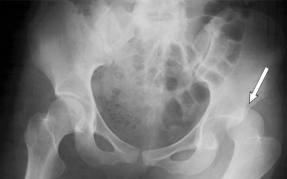
Anterior displacement of one vertebra over another can be seen on a lateral view of the spine. The displacement can be graded into four categories depending upon the severity of slip. Grade I spondylolisthesis means vertebral displacement up to 25 percent of the antero-posterior width of the lower vertebral body, whereas grade IV means the complete forward displacement of the affected vertebra. An oblique view of the spine may show defect in the pars interarticularis. In this view, in a normal vertebra, the pars interarticularis looks like a scottish dog. If the appearance is that of a scottish dog 'wearing a collar', the defect is in the isthmus (pars interarticularis), and the patient has a spondylolysis. If the head of the 'scottish dog' is separated from the neck, the patient has spondylolisthesis.
For a mild symptomless spondylolisthesis, no treatment is required. When symptoms are mild, they are adequately relieved by conservative methods, such as brace and spinal exercises. When symptoms are moderately severe or more, especially if these hamper the activity of the patient, an operation may be required.
Conservative methods consist of rest and external support to the affected segment followed by flexion exercises. The patient is advised to change his job to a physically less demanding one.
Operative methods consist of decompression of the compressed nerves if any, followed by fusion of the affected segments of the spine. This is commonly achieved by fusion between the transverse processes of adjacent vertebrae (inter- transverse fusion). Use of internal fixation devices like pedicular screws and rods has helped in early mobilisation of the patient.